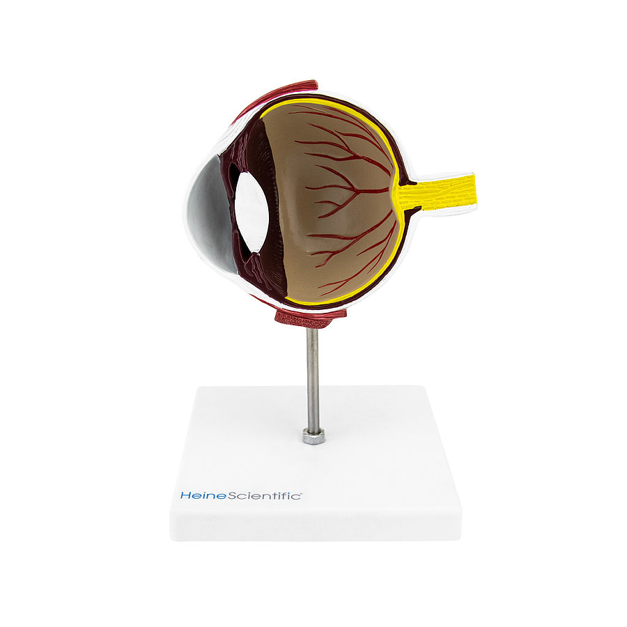
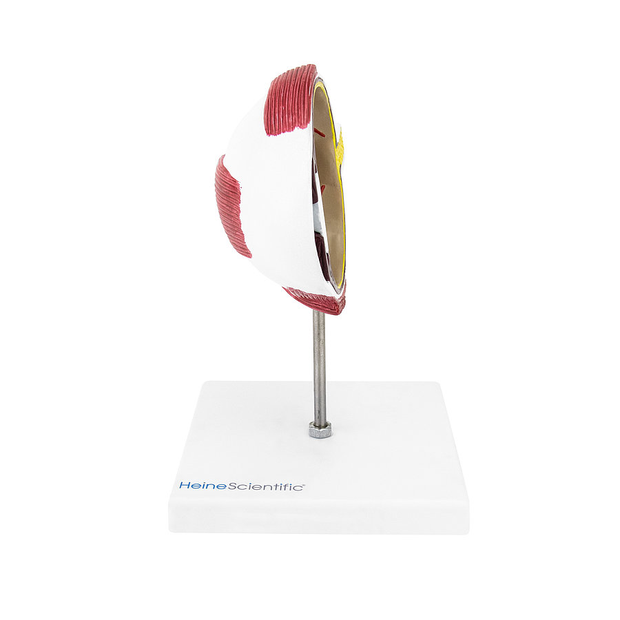
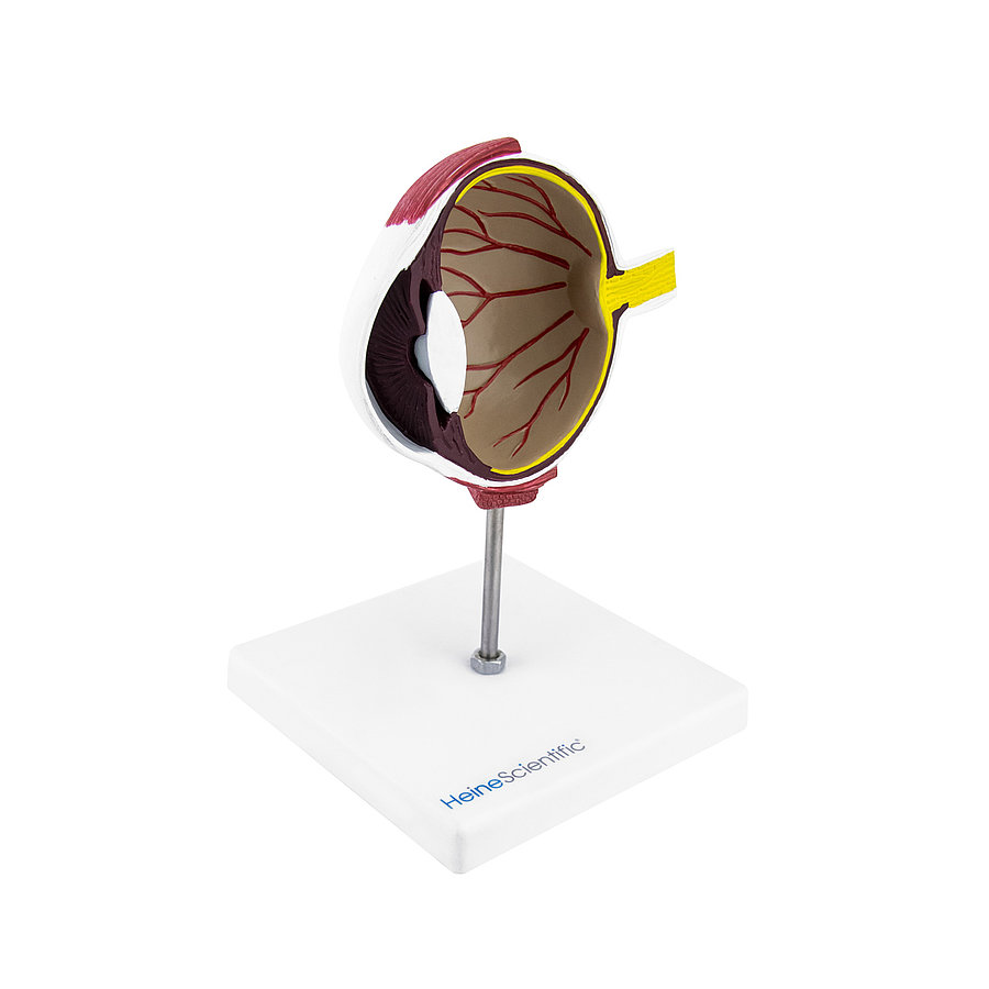


Cross-sectional model of a dog's eye
This canine eye model shows a cross section of the animal’s visual organ. The structure is easily recognisable, due to the colour accents and precise representation of individual parts. The model is securely held upright with a plastic base.
Visible Structures:
- Cornea (suggested)
- Anterior ocular chamber
- Iris
- Pupil
- Lens
- Vitreous body
- Retina
- Choroid
- Sclera
- Ciliary muscle
- Optic nerve
PRODUCT DETAILS
- Cross-sectional model of a dog’s eye
- For emphasising diagnoses and therapies
- Colour contrast for distinguishing between structures
- With white plastic base
- Base dimensions (L x W x H) : approx. 11.5 x 11.5 x 1 cm
- Model dimensions (L x W x H) : approx. 12 x 5 x 10 cm
Article Number: H191781 Category: SECTIONAL CUT ANATOMY MODELS